
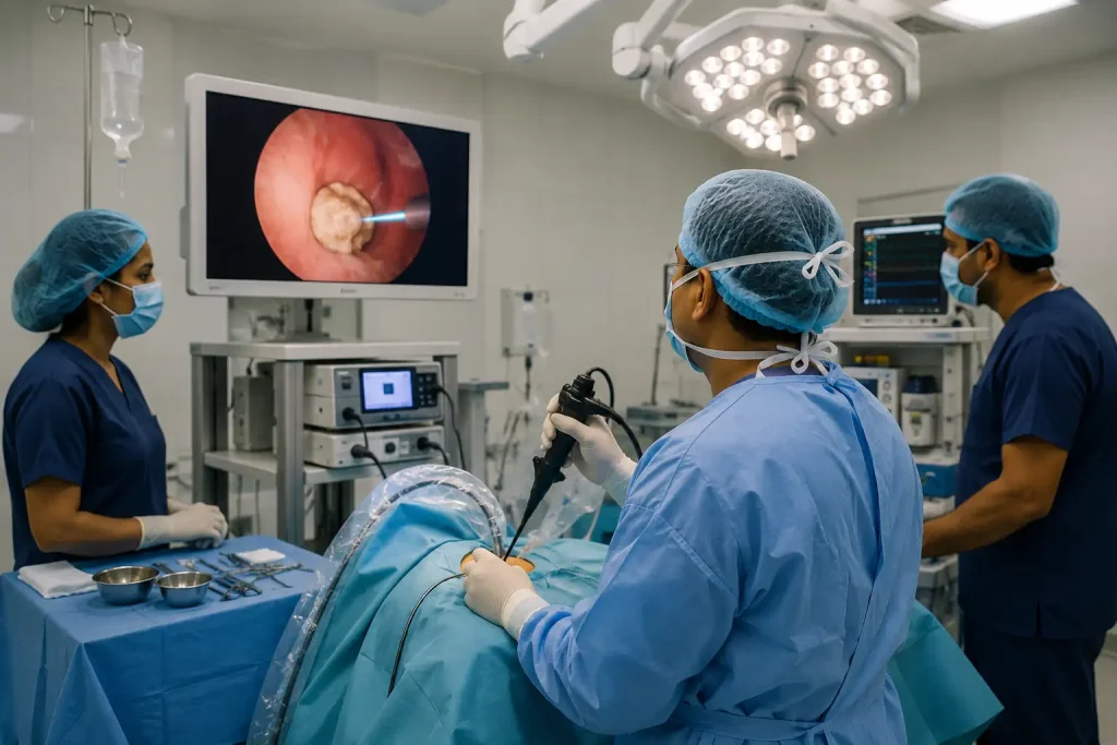
Urinary Stone Surgery: At a Glance
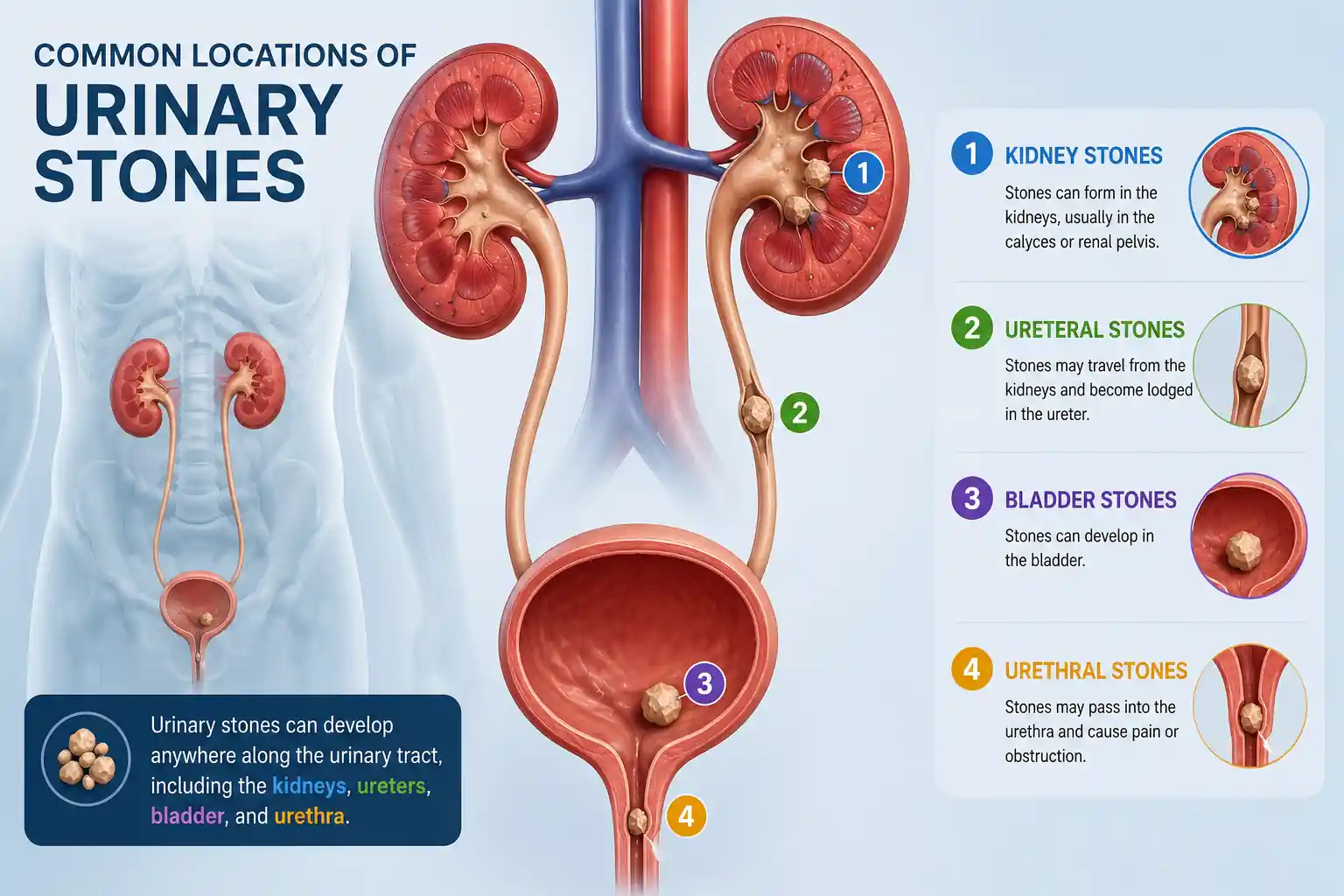
Urinary stone disease is one of the most common conditions affecting the urinary system, impacting millions of people worldwide each year. Stones can develop anywhere along the urinary tract—including the kidneys, ureters, bladder, or urethra—and may range from tiny crystals that pass unnoticed to larger stones capable of causing severe pain, urinary obstruction, infection, and even permanent kidney damage if left untreated.
Although many small stones can be managed with hydration, medication, and careful observation, larger or obstructive stones often require surgical intervention. Thanks to remarkable advances in urological technology, most urinary stones can now be treated using minimally invasive procedures that avoid the need for large surgical incisions. Modern techniques such as Laser Ureteroscopy (URS), Retrograde Intrarenal Surgery (RIRS), Percutaneous Nephrolithotomy (PCNL), and Endoscopic Bladder Stone Surgery offer excellent stone-free rates while reducing pain, shortening hospital stays, and allowing patients to return to normal life much sooner than traditional open surgery.
The most appropriate treatment depends on several important factors, including the stone’s size, location, composition, associated symptoms, kidney function, and overall health of the patient. A thorough clinical evaluation, supported by advanced imaging and laboratory investigations, enables the urologist to recommend a treatment plan tailored to each individual’s needs.
This comprehensive guide explains everything you need to know about urinary stone surgery—from understanding how stones form and recognizing early symptoms to diagnosis, modern treatment options, recovery, long-term prevention, and answers to the most frequently asked patient questions.
Quick Facts About Urinary Stone Surgery
| Procedure Time | Usually 30 minutes–2 hours |
|---|---|
| Anaesthesia | General or Spinal |
| Hospital Stay | Same day to 3 days |
| Recovery | 3 days–2 weeks (depending on procedure) |
| Success Rate | High with modern minimally invasive techniques |
| Return to Work | Usually within 3–14 days |
Understanding Urinary Stones
Urinary stones, also known as urinary tract stones or urolithiasis, are hard deposits of minerals and salts that develop within the urinary system. They form when certain substances naturally present in urine—such as calcium, oxalate, uric acid, or cystine—become highly concentrated and begin to crystallize. Over time, these tiny crystals stick together and gradually grow into stones of varying sizes.
Stones may develop in the kidneys, ureters, bladder, or urethra, and each location can produce different symptoms and require a different treatment approach. Some stones remain very small and pass naturally through the urine without causing significant discomfort. Others become lodged within the urinary tract, blocking the normal flow of urine and triggering severe pain, infection, or impaired kidney function.
Urinary stone disease affects people of all ages but is more common in adults between 30 and 60 years of age. Men generally have a higher risk than women, although the incidence among women has increased in recent years due to changing lifestyle and dietary patterns. Factors such as inadequate water intake, excessive salt consumption, obesity, metabolic disorders, recurrent urinary tract infections, and family history all contribute to an increased likelihood of stone formation.
If left untreated, larger stones may lead to complications such as hydronephrosis (swelling of the kidney), recurrent infections, reduced kidney function, or permanent kidney damage. Fortunately, advances in modern urology now allow most urinary stones to be treated safely using minimally invasive techniques with excellent long-term outcomes.
Signs and Symptoms You Should Never Ignore
The symptoms of urinary stones vary depending on the size, location, and movement of the stone. While some small stones may pass unnoticed, others can cause sudden and intense pain requiring urgent medical attention.
The most characteristic symptom is renal colic—a severe, wave-like pain that begins in the back or side below the ribs and may radiate toward the lower abdomen, groin, or genital area as the stone moves through the ureter. Many patients describe this as one of the most painful experiences they have ever had.
As the stone irritates the urinary tract, blood may appear in the urine, turning it pink, red, or brown. Some people experience burning during urination, increased urinary frequency, urgency, or difficulty passing urine. If the stone causes complete blockage or is associated with an infection, fever, chills, nausea, vomiting, and general weakness may develop. These symptoms require immediate medical evaluation.
It is important to remember that the intensity of pain does not always reflect the size of the stone. Even a small stone can cause severe discomfort if it becomes trapped in a narrow section of the ureter, while a larger stone may remain in the kidney for months with few or no symptoms.
Common Symptoms
- Severe pain in the back, side, or lower abdomen
- Pain radiating to the groin or genital area
- Blood in the urine (hematuria)
- Burning during urination
- Frequent or urgent urination
- Difficulty passing urine
- Cloudy or foul-smelling urine
- Nausea and vomiting
- Fever and chills (when infection is present)
Medical Alert: Seek immediate medical care if severe pain is accompanied by fever, chills, persistent vomiting, or an inability to pass urine.
What Causes Urinary Stones?
Urinary stones develop when the natural balance of water, minerals, and waste products in the urine is disrupted. Under normal conditions, urine contains substances that prevent crystals from forming. However, when urine becomes highly concentrated or contains excessive amounts of stone-forming minerals, tiny crystals begin to develop. Over time, these crystals join together, gradually forming stones that may increase in size and eventually block the urinary tract.
Although anyone can develop urinary stones, certain medical conditions, lifestyle habits, and genetic factors significantly increase the risk. Understanding these underlying causes is essential because successful treatment involves not only removing the existing stone but also preventing future recurrence.
One of the most common causes is inadequate fluid intake. When the body becomes dehydrated, urine becomes more concentrated, allowing minerals such as calcium, oxalate, and uric acid to crystallize more easily. This is particularly common in people living in hot climates, individuals who exercise heavily without replacing lost fluids, or those who simply do not drink enough water throughout the day.
Diet also plays an important role. A diet high in salt increases calcium excretion into the urine, while excessive consumption of animal protein may raise uric acid levels and lower urinary citrate, a natural inhibitor of stone formation. High intake of sugary drinks, processed foods, and obesity have also been associated with an increased risk of urinary stone disease.
Certain medical conditions—including gout, diabetes, hyperparathyroidism, recurrent urinary tract infections, inflammatory bowel disease, and some inherited metabolic disorders—can further increase stone risk. In addition, individuals with a family history of kidney stones are more likely to develop stones themselves, suggesting a significant genetic contribution.
Understanding these risk factors allows both patients and healthcare providers to develop personalized prevention strategies after successful treatment.
Common Risk Factors
- Drinking too little water
- Hot climate and excessive sweating
- High-salt diet
- Excessive animal protein intake
- Obesity
- Diabetes
- Gout
- Recurrent urinary tract infections
- Family history of urinary stones
- Certain metabolic disorders
- Previous urinary stones
- Sedentary lifestyle
Types of Urinary Stones
Not all urinary stones are the same. They differ in location, chemical composition, size, and hardness, and these differences influence both treatment decisions and long-term prevention strategies.
Understanding the exact type of stone helps doctors recommend the most appropriate surgical technique and advise patients on dietary and lifestyle changes that reduce the risk of recurrence.
Types of Urinary Stones by Location
Kidney Stones
Kidney stones develop inside one or both kidneys and are the most common form of urinary stone disease. Small stones may remain symptom-free for months or years, while larger stones can obstruct urine flow and cause severe pain when they move into the ureter.
Ureteric Stones
These stones become lodged in one of the ureters—the narrow tubes connecting the kidneys to the bladder. Because the ureter is relatively narrow, even small stones may cause intense renal colic and urinary obstruction.
Bladder Stones
Bladder stones usually develop when urine remains in the bladder for prolonged periods due to incomplete emptying. They are more common in older men with enlarged prostates but may also occur in patients with neurogenic bladder disorders or chronic urinary infections.
Urethral Stones
Urethral stones are relatively uncommon but can cause sudden difficulty passing urine, painful urination, and even complete urinary retention. Prompt medical evaluation is essential because complete blockage requires urgent treatment.
Types of Urinary Stones by Composition
The chemical composition of a urinary stone provides valuable information about why it formed and how future stones may be prevented.
Calcium Stones
Calcium stones account for nearly 80% of all urinary stones, making them the most common type. Most are composed of calcium oxalate, although calcium phosphate stones also occur.
They are often associated with dehydration, high sodium intake, excessive oxalate consumption, obesity, and certain metabolic disorders.
Uric Acid Stones
Uric acid stones develop when urine becomes persistently acidic. They are more common in individuals with gout, diabetes, obesity, or diets rich in red meat and seafood.
Unlike calcium stones, many uric acid stones can be prevented—or even dissolved—with medications that make the urine less acidic.
Struvite Stones
Struvite stones usually occur in patients with recurrent urinary tract infections caused by specific bacteria. These stones can grow rapidly and may eventually fill large portions of the kidney, forming staghorn calculi.
Because they are closely linked to infection, treatment generally requires complete stone removal together with appropriate antibiotic therapy.
Cystine Stones
Cystine stones are rare and result from an inherited genetic disorder known as cystinuria, in which excessive amounts of cystine are excreted in the urine.
Patients often develop recurrent stones from a young age and usually require lifelong preventive treatment, including aggressive hydration and specialized medications.
Stone Composition Comparison
| Stone Type | Most Common Cause | Approximate Frequency |
|---|---|---|
| Calcium | Dehydration, diet, metabolic disorders | ~80% |
| Uric Acid | Acidic urine, gout | 5–10% |
| Struvite | Urinary tract infection | 10–15% |
| Cystine | Genetic disorder | <2% |
How Are Urinary Stones Diagnosed?
Accurate diagnosis is the foundation of successful urinary stone treatment. Before recommending surgery, your urologist must determine the stone’s size, location, number, composition, and its effect on kidney function. This information guides the choice of treatment and helps identify any complications that require urgent intervention.
The evaluation begins with a detailed medical history and physical examination. Your doctor will ask about the onset and pattern of pain, urinary symptoms, previous stone episodes, dietary habits, medication use, and family history of urinary stones.
Laboratory investigations are then performed to assess kidney function, identify infection, and detect blood or crystals in the urine. Imaging studies provide precise information about the stone and remain essential for planning treatment.
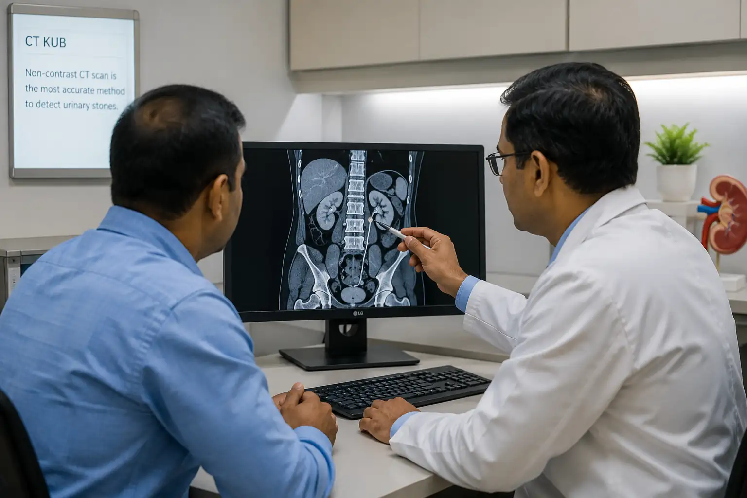
Among all imaging methods, Non-Contrast CT KUB (Computed Tomography of the Kidneys, Ureters, and Bladder) is considered the gold standard because it can detect almost all urinary stones with excellent accuracy. Ultrasound is also widely used, particularly in pregnant women and for follow-up evaluations, while plain X-rays may help monitor certain types of stones.
Common Diagnostic Tests
- Medical history and physical examination
- Urinalysis
- Urine culture
- Blood tests
- Kidney function tests
- Ultrasound
- CT KUB (Gold Standard)
- X-ray KUB (selected cases)
When Is Urinary Stone Surgery Necessary?
Not every urinary stone requires surgery. Many small stones pass naturally with adequate hydration, pain management, and appropriate medication. However, surgical treatment becomes necessary when a stone is unlikely to pass on its own or when it begins to threaten kidney function or the patient’s overall health.
Your urologist will recommend surgery based on several important factors, including the stone’s size, location, degree of obstruction, associated symptoms, presence of infection, and response to conservative treatment.
In general, larger stones are much less likely to pass spontaneously and are more likely to require minimally invasive intervention. Similarly, stones causing persistent pain, repeated urinary infections, bleeding, or impaired kidney drainage should not be left untreated.
Modern minimally invasive procedures allow these stones to be removed safely while minimizing discomfort and promoting faster recovery.
Surgery Is Usually Recommended When
- The stone is too large to pass naturally.
- Severe pain persists despite medication.
- Urine flow is blocked.
- Kidney function is threatened.
- There is recurrent urinary tract infection.
- Blood in the urine continues.
- Conservative treatment has failed.
- Stones continue to enlarge over time.
- Recurrent stone episodes significantly affect quality of life.
Clinical Tip: Early intervention often prevents serious complications such as permanent kidney damage, recurrent infections, and emergency hospitalization.
Modern Treatment Options for Urinary Stones
The treatment of urinary stones has advanced significantly over the past two decades. In the past, many patients required open surgery involving large incisions, prolonged hospitalization, and lengthy recovery periods. Today, thanks to remarkable developments in endourology and laser technology, most urinary stones can be treated using minimally invasive procedures that are safer, less painful, and highly effective.
The most appropriate treatment depends on several factors, including the stone’s size, location, composition, degree of obstruction, symptoms, and overall kidney function. After reviewing your imaging studies and laboratory results, your urologist will recommend the option that offers the highest chance of complete stone removal while minimizing risks and recovery time.
Not every patient requires surgery. Small stones may pass naturally with hydration, pain relief, and medications. However, larger stones or those causing persistent pain, infection, or obstruction usually require procedural intervention.
Treatment Options Available Today
Modern management of urinary stones may include:
- Conservative treatment (observation and hydration)
- Medical Expulsive Therapy (MET)
- Extracorporeal Shock Wave Lithotripsy (ESWL)
- Laser Ureteroscopy (URS)
- Retrograde Intrarenal Surgery (RIRS)
- Percutaneous Nephrolithotomy (PCNL)
- Endoscopic Bladder Stone Surgery
- Open or Laparoscopic Surgery (rarely required)
Today, more than 90% of urinary stones can be treated without traditional open surgery, thanks to advances in minimally invasive techniques.
Conservative Treatment (Observation & Medication)
Not every urinary stone requires an operation. Small stones—particularly those measuring less than 5 mm—often pass naturally through the urinary tract.
Your doctor may recommend conservative management if:
- The stone is small.
- Pain is controlled.
- There is no infection.
- Kidney function remains normal.
- The urinary tract is not completely blocked.
Treatment usually includes pain medication, increased fluid intake, and medications that relax the ureter, making it easier for the stone to pass naturally.
During this period, regular follow-up is important to ensure the stone progresses safely without causing complications.
Extracorporeal Shock Wave Lithotripsy (ESWL)
ESWL is a completely non-invasive treatment that uses high-energy shock waves to break urinary stones into smaller fragments.
The procedure does not require an incision or insertion of instruments into the urinary tract. Instead, shock waves generated outside the body are precisely focused onto the stone using ultrasound or X-ray guidance.
After fragmentation, the tiny pieces pass naturally in the urine over several days or weeks.
Although ESWL remains an effective treatment for selected patients, it is not suitable for every type of stone. Larger, harder, or lower ureteric stones often respond better to endoscopic laser procedures.
ESWL is commonly recommended for:
- Small kidney stones
- Selected upper ureteric stones
- Patients preferring a completely non-invasive option
Advantages
- No incision
- Outpatient procedure
- Quick recovery
- Minimal discomfort
Laser Ureteroscopy (URS)
Laser Ureteroscopy (URS) is considered the gold standard treatment for most ureteric stones and selected kidney stones.
Unlike traditional surgery, URS is performed without any external incision. A thin, flexible endoscope is gently inserted through the urethra, passed into the bladder, and advanced into the ureter until the stone is identified.
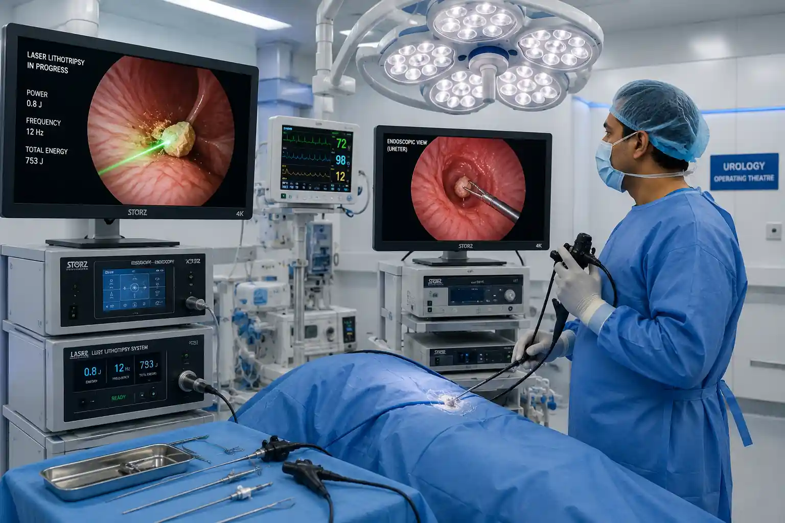
Once the stone is visualized, a Holmium:YAG Laser or Thulium Fiber Laser (TFL) is used to fragment it into tiny pieces. Small fragments may be removed using miniature baskets, while microscopic particles pass naturally in the urine.
If swelling is expected, a temporary Double-J (DJ) ureteral stent may be inserted to maintain urine drainage during healing. This stent is usually removed during a short outpatient visit.
Because URS causes minimal tissue trauma, patients generally experience less pain, little bleeding, and a much faster recovery than with open surgery.
URS is ideal for
- Ureteric stones
- Small to medium kidney stones
- Stones causing urinary obstruction
- Persistent severe pain
- Failed conservative treatment
Benefits
- No external incision
- High success rate
- Minimal bleeding
- Short hospital stay
- Fast recovery
- Excellent cosmetic outcome
Retrograde Intrarenal Surgery (RIRS)
Retrograde Intrarenal Surgery (RIRS) represents one of the most advanced techniques available for treating kidney stones.
Using an ultra-flexible digital ureteroscope, the surgeon reaches almost every part of the kidney through the body’s natural urinary tract. Once the stone is located, a high-powered laser breaks it into dust-like particles that are removed or passed naturally.
Because RIRS requires no skin incision, postoperative discomfort is usually mild, and recovery is remarkably fast.
RIRS is recommended for
- Kidney stones under 2 cm
- Multiple kidney stones
- Difficult-to-reach stones
- Patients requiring scar-free surgery
Advantages
- Completely incision-free
- Excellent stone clearance
- Minimal postoperative pain
- Short hospitalization
- Rapid recovery
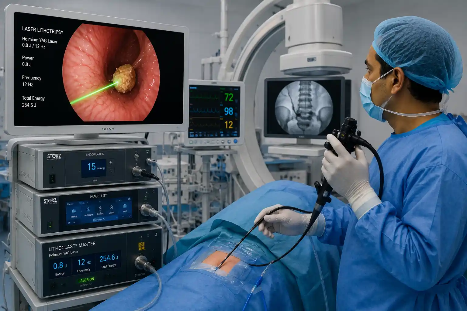
Percutaneous Nephrolithotomy (PCNL)
For large, complex, or staghorn kidney stones, Percutaneous Nephrolithotomy (PCNL) remains the gold standard surgical treatment.
Unlike URS or RIRS, PCNL involves creating a very small incision—typically about 1 cm—in the patient’s back. Through this opening, the surgeon gains direct access to the kidney using specialized instruments.
The stone is fragmented using ultrasonic, pneumatic, or laser technology before being removed through the access tract.
Although PCNL is slightly more invasive than other minimally invasive procedures, it offers the highest stone-free rate for patients with large stone burdens while still avoiding the extensive trauma associated with traditional open surgery.
PCNL is best for
- Stones larger than 2 cm
- Staghorn calculi
- Multiple large kidney stones
- Complex stone disease
Benefits
- Highest success rate
- Excellent for large stones
- Fewer repeat procedures
- Kidney preservation
- Faster recovery than open surgery
Endoscopic Bladder Stone Surgery (Cystolitholapaxy)
Bladder stones are usually treated using an endoscopic procedure known as cystolitholapaxy.
A cystoscope is inserted through the urethra into the bladder, allowing the surgeon to visualize the stone directly. Laser or mechanical devices fragment the stone, and the pieces are removed through the cystoscope.
If an enlarged prostate or another cause of bladder outlet obstruction contributed to stone formation, your doctor may recommend treating that underlying condition as well.
Benefits
- No external incision
- Excellent success rate
- Minimal discomfort
- Same-day discharge in many patients
- Quick recovery
Comparison of Modern Urinary Stone Procedures
| Procedure | Best For | Incision | Hospital Stay | Recovery |
|---|---|---|---|---|
| Conservative Treatment | Small stones | ❌ | None | Natural passage |
| ESWL | Small kidney stones | ❌ | Same day | 2–7 days |
| URS | Ureteric stones | ❌ | Same day–1 day | 3–5 days |
| RIRS | Kidney stones (<2 cm) | ❌ | 1 day | 5–7 days |
| PCNL | Large kidney stones (>2 cm) | ✅ Small | 2–3 days | 1–2 weeks |
| Cystolitholapaxy | Bladder stones | ❌ | Same day | 2–5 days |
Benefits of Minimally Invasive Urinary Stone Surgery
Modern minimally invasive surgery has transformed the treatment of urinary stones by reducing surgical trauma while maintaining outstanding success rates. Compared with traditional open surgery, these techniques allow patients to recover more quickly and return to normal activities sooner.
For most patients, minimally invasive procedures provide a safer, more comfortable experience with excellent long-term outcomes.
Key Benefits
- No or very small incision
- Less postoperative pain
- Minimal blood loss
- Lower infection risk
- High stone-free success rates
- Short hospital stay
- Faster recovery
- Better cosmetic results
- Preservation of kidney function
- Earlier return to work and daily life
Clinical Insight: Choosing the right procedure depends on the size, location, and composition of the stone, as well as your overall health. Your urologist will recommend the treatment option that offers the best balance of safety, effectiveness, and long-term success.
Preparing for Urinary Stone Surgery
Successful urinary stone surgery begins long before you enter the operating room. Careful preparation helps reduce surgical risks, improves treatment outcomes, and supports a faster recovery. Before recommending surgery, your urologist will perform a comprehensive evaluation to understand your overall health, assess the complexity of the stone, and select the most appropriate surgical technique.
During your consultation, your doctor will discuss your symptoms, previous stone history, current medications, allergies, and any underlying medical conditions such as diabetes, hypertension, heart disease, or bleeding disorders. This information is essential for planning a safe procedure and minimizing potential complications.
Imaging studies, including CT scans or ultrasound examinations, are carefully reviewed to determine the exact size, location, and number of stones. Laboratory investigations help identify urinary tract infections, assess kidney function, and ensure you are medically fit for anesthesia.
If you are taking blood-thinning medications, your healthcare provider may recommend temporarily adjusting them before surgery. However, these medications should never be stopped without professional medical advice. You will also receive instructions about fasting before anesthesia and when to stop eating or drinking before the procedure.
Proper preparation not only improves surgical safety but also gives patients a clear understanding of what to expect before, during, and after treatment.
Before Surgery, Your Doctor May Recommend
- Complete medical evaluation
- Urinalysis and urine culture
- Blood investigations
- Kidney function tests
- CT KUB or ultrasound
- ECG (when indicated)
- Chest X-ray (selected patients)
- Medication review
- Temporary adjustment of blood-thinning medications (if required)
- Fasting before anesthesia
 What Happens During Urinary Stone Surgery?
Although each surgical technique differs slightly, the overall objective remains the same—to remove the stone safely while preserving normal urinary tract function and minimizing damage to surrounding tissues.
On the day of surgery, you will be admitted to the hospital and prepared by the nursing and anesthesia teams. After confirming your medical information and reviewing your imaging studies, anesthesia will be administered. Depending on the planned procedure, this may be general anesthesia or spinal anesthesia, ensuring that you remain comfortable throughout the operation.
For minimally invasive procedures such as Laser Ureteroscopy (URS) and Retrograde Intrarenal Surgery (RIRS), the surgeon inserts a fine endoscope through the urethra into the bladder and then carefully advances it into the ureter or kidney. Once the stone is located, advanced laser technology is used to fragment it into tiny pieces that are removed or allowed to pass naturally through the urine.
For larger kidney stones requiring Percutaneous Nephrolithotomy (PCNL), a small incision—typically about one centimeter—is made in the patient’s back to provide direct access to the kidney. Specialized instruments are then used to fragment and remove the stone safely.
In some cases, a temporary Double-J (DJ) ureteral stent is placed at the end of surgery. This flexible tube helps maintain urine drainage, reduces swelling, and promotes healing. It is usually removed during a brief outpatient visit after several days or weeks.
Depending on the complexity of the stone and the chosen procedure, surgery generally lasts between 30 minutes and 2 hours.
How Long Does Recovery Take?
One of the greatest advantages of modern minimally invasive urinary stone surgery is the speed of recovery. Because these procedures cause significantly less tissue trauma than traditional open surgery, most patients can begin walking within a few hours and return home much sooner.
It is normal to experience mild discomfort, burning during urination, or a small amount of blood in the urine for the first few days. These symptoms usually improve quickly as the urinary tract heals. Patients with a temporary ureteral stent may notice urinary urgency, increased frequency, or mild flank discomfort until the stent is removed.
Recovery time varies according to the type of surgery, the size and location of the stone, and the patient’s overall health. Most patients resume light daily activities within several days, while more physically demanding work may require additional recovery time.
Following your surgeon’s instructions carefully and attending scheduled follow-up appointments are essential for ensuring complete healing and confirming that all stone fragments have been removed.
Recovery Timeline
| Time After Surgery | What You Can Expect |
|---|---|
| First 24 Hours | Mild discomfort, early walking encouraged |
| 2–3 Days | Improved pain, gradual return to normal activities |
| 1 Week | Most patients feel significantly better |
| 2 Weeks | Many patients resume normal routines |
| 4–6 Weeks | Complete recovery in most cases |
Recovery time varies depending on the procedure performed and each patient’s individual condition.
Post-Operative Care: Tips for a Smooth Recovery
The success of urinary stone surgery depends not only on the procedure itself but also on proper postoperative care. Following your surgeon’s recommendations carefully reduces the risk of complications and supports a quicker, smoother recovery.
Hydration is particularly important during the recovery period. Drinking plenty of water helps flush out tiny stone fragments, reduces irritation within the urinary tract, and supports normal kidney function. Unless otherwise advised by your doctor, aim to drink enough fluids throughout the day to keep your urine light yellow or nearly clear.
Take all prescribed medications exactly as directed. These may include pain relievers, antibiotics, or medications to reduce ureteral spasms. Completing the full course of antibiotics, when prescribed, is especially important to prevent infection.
Although heavy lifting and strenuous physical activity should be avoided during the early recovery period, gentle walking is strongly encouraged. Walking improves blood circulation, reduces the risk of blood clots, and promotes faster healing.
If a temporary DJ stent has been inserted, you may experience mild urinary discomfort until it is removed. This is usually temporary and resolves soon after stent removal.
Recovery Tips
For the best possible recovery:
- Drink 2.5–3 liters of water daily unless advised otherwise.
- Take all prescribed medications.
- Walk regularly.
- Avoid heavy lifting until approved by your surgeon.
- Eat a balanced, nutritious diet.
- Attend all scheduled follow-up appointments.
- Complete any prescribed antibiotic course.
- Contact your doctor if symptoms worsen.
Diet After Urinary Stone Surgery
Diet plays a vital role in both recovery and the prevention of future urinary stones. Surgery removes existing stones, but maintaining healthy dietary habits helps reduce the risk of recurrence and supports long-term urinary tract health.
Adequate hydration remains the single most important dietary recommendation. Drinking sufficient water helps dilute the urine, preventing minerals from becoming concentrated enough to form new stones. Most patients should aim to drink 2.5–3 liters of fluids daily, unless otherwise advised due to another medical condition.
A balanced diet rich in fresh fruits, vegetables, whole grains, and lean proteins supports overall kidney health. Limiting excessive salt intake is particularly important because sodium increases calcium excretion into the urine, making stone formation more likely.
Patients with specific types of stones may receive individualized dietary recommendations. For example, those with uric acid stones may be advised to reduce excessive red meat intake, while patients prone to calcium oxalate stones may benefit from moderating foods naturally high in oxalates. Rather than following restrictive diets, the goal is to establish sustainable eating habits that promote lifelong urinary health.
Healthy Dietary Recommendations
- Drink plenty of water throughout the day.
- Reduce excessive salt intake.
- Eat more fresh fruits and vegetables.
- Choose whole grains whenever possible.
- Consume moderate amounts of lean protein.
- Limit sugary soft drinks.
- Maintain a healthy body weight.
- Follow personalized dietary advice based on your stone type.
Healthy lifestyle changes after surgery are just as important as the surgery itself. They help protect your kidneys, reduce the risk of future stones, and support long-term urinary health.
Life After Urinary Stone Surgery: Long-Term Follow-up and Prevention
Successfully removing a urinary stone is only the first step toward better urinary health. Without addressing the underlying causes, new stones can develop months or even years later. Studies have shown that urinary stones have a relatively high recurrence rate, particularly in individuals who do not make appropriate lifestyle and dietary changes after treatment.
Long-term prevention focuses on reducing the concentration of stone-forming minerals in the urine while identifying and managing any medical conditions that contribute to stone formation. Your urologist may recommend regular follow-up visits, urine analysis, blood tests, or imaging studies depending on the type of stone you had and your individual risk factors.
Hydration remains the cornerstone of prevention. Producing an adequate volume of urine each day significantly reduces the likelihood of crystals forming inside the urinary tract. In addition, maintaining a balanced diet, controlling body weight, staying physically active, and following personalized dietary recommendations can dramatically lower the chance of recurrence.
Patients who have experienced recurrent stones or have inherited metabolic disorders may require additional medical therapy and closer long-term monitoring.
How to Prevent Future Urinary Stones
Although urinary stones cannot always be prevented, adopting healthy lifestyle habits can significantly reduce your risk of developing another stone.
Drink Plenty of Water
Maintaining good hydration is the most effective preventive measure. Most adults should aim to drink enough fluids to produce at least 2–2.5 liters of urine daily, unless otherwise advised by their doctor.
Reduce Salt Intake
A diet high in sodium increases calcium excretion in the urine, making calcium stone formation more likely. Reducing processed foods and avoiding excessive table salt can help lower this risk.
Eat a Balanced Diet
Fresh fruits, vegetables, whole grains, and moderate amounts of lean protein support kidney health. Avoid overly restrictive diets unless specifically recommended by your healthcare provider.
Maintain a Healthy Weight
Obesity and metabolic syndrome are associated with an increased risk of urinary stone disease. Regular exercise and healthy eating habits contribute to long-term prevention.
Follow Personalized Medical Advice
Patients with calcium, uric acid, cystine, or infection-related stones may require specific dietary modifications or medications. Your prevention plan should always be tailored to your stone type and medical history.
🥗 Lifestyle Tips for Long-Term Stone Prevention
- Drink water consistently throughout the day.
- Limit excessive salt intake.
- Maintain a healthy body weight.
- Stay physically active.
- Avoid excessive sugary beverages.
- Follow your prescribed medications.
- Attend regular follow-up appointments.
- Seek medical evaluation if symptoms return.
Possible Risks and Complications of Urinary Stone Surgery
Modern minimally invasive urinary stone surgery is considered very safe and highly successful. However, as with any surgical procedure, certain risks and complications may occur. Fortunately, serious complications are uncommon, especially when surgery is performed by an experienced urologist in an appropriately equipped medical facility.
Most complications, if they occur, are temporary and can be managed effectively with prompt medical attention.
Potential complications may include:
- Mild blood in the urine for a short period
- Temporary burning during urination
- Urinary tract infection
- Fever
- Pain related to a temporary ureteral stent
- Incomplete stone clearance requiring additional treatment
- Rare injury to the urinary tract
- Very rare bleeding requiring intervention
Your surgeon will discuss these risks with you before surgery and explain the measures taken to minimize them.
When Should You Visit the Emergency Department?
Seek immediate medical attention if you experience any of the following after urinary stone surgery:
- High fever (above 38.5°C) or shaking chills
- Severe or worsening flank pain that is not relieved by prescribed medication
- Inability to pass urine
- Heavy bleeding or large blood clots in the urine
- Persistent nausea and vomiting preventing fluid intake
- Difficulty breathing or chest pain
- Severe abdominal swelling
- Sudden decrease in urine output
- Signs of an allergic reaction to medication
Do not delay seeking medical care if any of these warning signs develop. Early treatment can prevent serious complications and protect kidney function.
Frequently Asked Questions (FAQ)
Is urinary stone surgery painful?
Modern minimally invasive procedures are performed under general or spinal anesthesia, so you will not feel pain during the operation. Mild discomfort afterward is common but is usually well controlled with prescribed pain medication.
How long will I stay in the hospital?
Many patients undergoing URS, RIRS, or bladder stone surgery are discharged the same day or within 24 hours. Patients undergoing PCNL may stay in the hospital for two to three days depending on their recovery.
How soon can I return to work?
Most patients return to office-based work within 3–7 days after minimally invasive surgery. Jobs involving heavy physical activity may require a longer recovery period.
Will the stone come back after surgery?
Surgery removes existing stones but does not eliminate the underlying tendency to form new stones. Maintaining adequate hydration, following dietary recommendations, and attending follow-up appointments can significantly reduce the risk of recurrence.
Will I need a ureteral stent?
Not every patient requires a stent. When used, a Double-J (DJ) stent is temporary and is usually removed during a short outpatient visit.
Can urinary stones damage my kidneys?
Yes. Untreated stones that block urine flow or cause recurrent infections may eventually impair kidney function or lead to permanent kidney damage.
Which surgery is best for urinary stones?
There is no single procedure that is best for every patient. The ideal treatment depends on the stone’s size, location, composition, symptoms, and your overall health. Your urologist will recommend the most appropriate option after evaluation.
How can I reduce my risk of developing another stone?
Drinking enough water, limiting salt intake, maintaining a healthy weight, eating a balanced diet, and following your doctor’s advice based on your stone type are the most effective preventive strategies.
Can all urinary stones be treated without open surgery?
In most cases, yes. Advances in endoscopic and laser technology mean that the majority of urinary stones can now be treated using minimally invasive techniques without the need for traditional open surgery.
When should I contact my doctor after surgery?
You should contact your healthcare provider immediately if you develop fever, severe pain, heavy bleeding, inability to pass urine, persistent vomiting, or any other concerning symptoms during recovery.
Consult a Specialist Today
If you are experiencing persistent flank pain, blood in your urine, recurrent urinary tract infections, or have been diagnosed with a urinary stone, early consultation with an experienced surgeon can help prevent complications and ensure timely treatment.
Modern minimally invasive techniques offer excellent success rates with less pain, smaller or no incisions, shorter hospital stays, and faster recovery. Seeking professional evaluation at the right time can make a significant difference in both treatment outcomes and long-term kidney health.
Medical Disclaimer
This article is intended for educational and informational purposes only and should not be considered a substitute for professional medical advice, diagnosis, or treatment. Every patient is unique, and treatment recommendations vary depending on individual medical history, examination findings, and diagnostic results. Always consult a qualified healthcare professional for personalized medical advice. If you experience severe pain, fever, difficulty passing urine, heavy bleeding, or any medical emergency, seek immediate medical attention.


